Pages

Aug 23, 2010

Kill the shine.


Do you ever get annoyed by how shiny molecules are? Every picture of a molecule I see shows a shiny shiny molecule, like it was made of alien-space-age-nasa-plastic. Here I show you how to kill that shine, making a dull earthlier look.

Easy to do: decrease specularity value to 0.08.


1. Open Deja vu. Select the 'Light' tab. Select 'Light Colors' box.


2. Set the drop down menu to "light 1- specular"


3. Adjust the Value from 80 (default) to 10 or below.


There's a lot of cool effects to be achieved by adjusting the values and colors of the various lights in the 'Light Colors' menu. Poke around to get a unique look.

Bonus: when you have a look you want to save, Bookmark it! In Deja vu, select 'Bookmarks' tab and click 'Save Representation'.

Aug 20, 2010

Calculate Volume

Quick: what is the volume of this molecular suface?

Here I present a simple method for calculating the volume of a geometric object in PMV.
1. Open Vision.
2. Load the Visualization Library.

3. Drag down the 'PolyhedronVolumeArea' node from the mapper menu and the 'Choose Geom' node from the filter menu in the Visualization library. Drag down the 'PMV Viewer' node in the PMV library. Drag down the 'PopUpMsg' from the Standard Library.
4. Arrange them: PMV Viewer to Choose Geom to PolyVolArea to PopUpMSG. See picture below:
Be sure to choose the geometry you'd like to calculate the volume for.
Here, I have chosen the molecular surface.


5. The volume will appear in the Python IDLE Shell.
Note: units are in Angstroms.

Measure Distance in PMV

Antibody molecule 1igt, with a wingspan of 152.411 Angstroms, or 15.2 nanometers.

1. Under 'Picking Commands', select 'measureDistanceGC'.
The 'Picking Commands' icon is a pointing hand in blue.

2. Select the two atoms you wish to measure the distance between.


Note: Units are Angstoms.

Aug 19, 2010

Ambient Occlusion in PMV

Antibody 1igt with Ambient Occlusion from PMV by Jon Huntoon

From wikipedia: "Ambient occlusion is a shading method used in 3D computer graphics which helps add realism to local reflection models by taking into account attenuation of light due to occlusion. Ambient occlusion attempts to approximate the way light radiates in real life, especially off what are normally considered non-reflective surfaces.
Unlike local methods like Phong shading, ambient occlusion is a global method, meaning the illumination at each point is a function of other geometry in the scene. However, it is a very crude approximation to full global illumination. The soft appearance achieved by ambient occlusion alone is similar to the way an object appears on an overcast day."

To achieve this sophisticated look is mostly unattainable in other viewing software. But PMV makes it painless. Here's how:

1. Open Deja Vu
2. Select Ambient Occlusion
3. Select the geometries you'd like to have ambient occlusion applied to and then click 'Add'.
4. Select cavity darkness and overall brightness (I left these settings default).
5. Click Compute.


That's all. Sit back and watch the computations chug away. My rendering took about 2 minutes on a new iMac. The other cool thing about this is that once the shadows are calculated, they remain. You don't need to recalculate every time the camera moves like you would in other 3D viewers. Have fun!

Here's what a secondary structure looks like with ambient occlusion turned on.
Ambient occlusion on secondary structure.
Note: the stick-and-balls didn't get rendered.


BONUS: In Deja Vu, select 'Scene Antialiasing' and increase the value to at least 8 for cleaner edges.


-Jon Huntoon

Jun 17, 2010

Distinct Molecular Surfaces Per Chain


How you decide to distinguish between chains in a molecule is really up to taste, but I think there is room for improvement in legibility. Most scientists distinguish chains by color. I do too. But we can go farther, and perhaps, do better.

By default, molecular surfaces are calculated with disregard for individual chains:

This is actually more true to 'real life' or 'in situ'. The water molecules defining the surface of a protein have no regard for chains. They simply pass over. But we operate 'in silico', and afforded the opportunity to distinguish with clarity between the molecular surfaces of chains. Perhaps for the better in the end for discovery- or at least another arrow in our quiver.

Here is a side by side comparison:


I think it is clear from the screen shots above that calculating the molecular surface per chain can be a positive augmentation of the representation.

So, without further adieu, I present the simple trick:

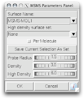
1. While in the computeMSMS popup menu, de-select the 'Per Molecule' check box.

2. In that same menu, be sure to rename the 'Surface Name' for each chain.


That's it!

Another quick tip is that you can reach the ComputeMSMS menu by right-clicking the radio buttons in dashboard:


Enjoy!

JON HUNTOON

Jun 7, 2010

Aligning Small Molecules

From there:  to here:

Aligning two small molecules is useful and necessary when studying the similarities of form and character of two distinct molecules. Here's one way of doing that in PMV.

1. Select COMPUTE--> SUPERIMPOSE ATOM PAIRS.


2. Select ADD PAIRS.


3. Turn the  BY STRING mode on. You could do this by picking atoms by hand as well. But I find the selecting by string is faster if your molecules have enough similar points (3).


4. The two molecules that I am aligning are methionine and glutamate. I know that they share at least their backbone atoms and the beta carbon "CB". All I need is 3 atoms for an alignment, so I just type "backbone" under the ATOM heading for the REFERENCE and MOBILE nodes. Note that the reference molecule will stay put, while the mobile molecule will be aligned onto the reference molecule, as you might expect.


Also note that by clicking SHOW you will see little blue and red stars over the reference and mobile atoms.


Click OKAY.

5.  Now click CREATE PAIRS  in the superimpose window.


 6. Your molecules should be aligned and you can  DISMISS the "pairwise superimposition" window.


Have fun, 
JON

Apr 27, 2010

Making David Goodsell like illustrations.


David Goodsell as you may know, is an exceptional artist and brilliant communicator. His watercolors have been published in magazines, journals, and books (The Machinery of LifeBionanotechnology: Lessons from Nature) and hung up in galleries and exhibits around the world. He writes a monthly column for the Protein Data Bank (PDB) called 'Molecule of the Month'

Lucky us, PMV has an auto 'David Goodsell' feature. 


1. Firstly, there is the "DG Colors" under the color menu. This coloring is similar to atomic but the intensity of the red and blue on oxygens and nitrogen respectively corresponds to the charge of the residue upon which the atom resides. For example, a highly negatively charge oxygens on glutamate will be deep red, where as the less negatively charged oxygens on threonine will be pink. Conversely, the strongly positive charged nitrogen upon the lysine will be dark blue while the less positive nitrogens on histidine are sky blue.  A clever coloring scheme that incorporate charge and atom property is one way in which the David Goodsell style conveys so much information without becoming too busy. 


2. Now that your models are colored according the custom DG scheme, it is time to emulate the subtle beauty of his water colors. Turn on the 'cartoon mode'.
adjust if you wish:


3. Now you are officially David Goodsell-esque. The last recommendation I have is to increase the anti-aliasing to at least 4. Beyond 8, the anti-aliasing tends to suck too much CPU power. 


ENJOY!
 
Powered by Blogger